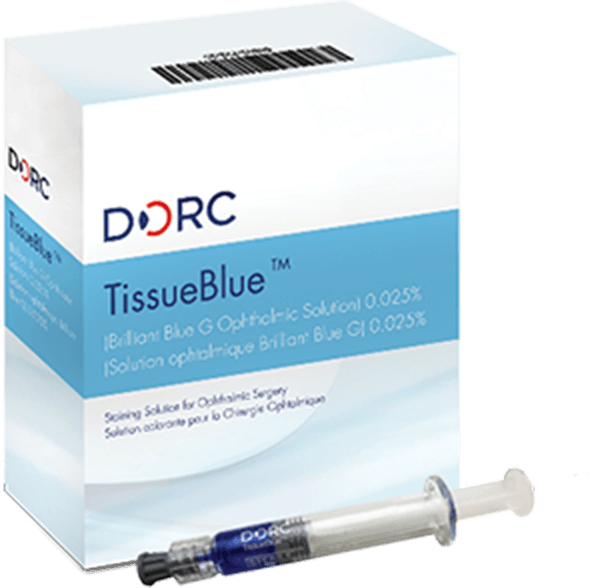
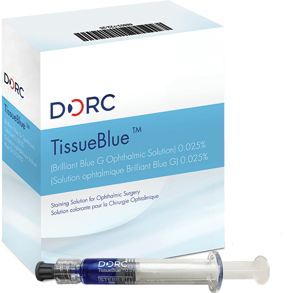
FDA-approved TissueBlue™
is ready for you.
(Brilliant Blue G Ophthalmic Solution) 0.025%
The ONLY FDA-approved selective stain for the ILM. Easy open-and-inject application from a pre-filled, sterile syringe.
*Sample available to registered US physicians only. Samples subject to availability.
Confidence. Consistency. Convenience.
Confidence of
superior purity1
FDA-approved, pharmaceutical-grade dye.
Consistency
& Convenience
Pre-filled, sterile syringe eliminates need to mix or source via compounding pharmacies.
PROVEN SAFETY†
Trusted for ILM staining in over 350,0002 procedures worldwide.
†Marketed as ILM Blue Outside US since 2010.
References
1. Data on file – Results of HPLC purity tests performed on samples of compounded BBG dyes available in the U.S.
2. Total DORC Global Sales data for ILM Blue since launch – available on file.
Watch clinical videos of ILM Blue† to see it in action.
Ma première chirurgie du trou maculaire avec pelage de la MLI en utilisant Tissueblue™ par Arshad Khanani, MD, USA.
Réussir la coloration sélective et le pelage MLI avec Tissueblue™ par Brian Joondeph, MD, USA
Asheesh Tewari, MD (Michigan, USA) réalise l'une de ses premières opérations avec Tissueblue.
Démonstration de chirurgie avec la « ILM Blue »
Coloration efficace avec Tissueblue™ Coloration de MLI
Vidéo comparative -
Tissueblue avec PEG et BBG sans PEG
†TissueBlue has been marketed as ILM Blue Outside US since 2010.
Request a free TissueBlue sample* today!

FDA approval of TissueBlue is great for U.S. retina specialists who have long-awaited an FDA-approved ILM dye that combines a very strong safety profile and excellent staining characteristics. This is a very welcome development.
Read published articles about TissueBlue.
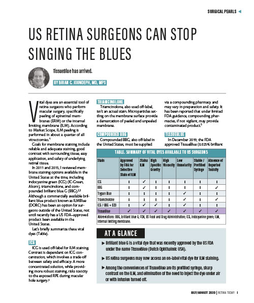
Among the conveniences of TissueBlue are its prefilled syringe, sharp contrast on the ILM, and elimination of the need to inject the dye under air or with infusion turned off.
(source: Retina Today, July/August 2020.)
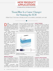
(source: Retinal Physician, June 2020)
Animal studies have shown the dose-dependent toxicity of indocyanine green, especially in case of diffusion into the subretinal space, where it can potentially cause severe retinal damage. In fact, because of the altered structure of the neuroretina, iatrogenic retinal breaks may allow indocyanine green to reach the sub-retinal space. In contrast, the safety profile of brilliant blue G is excellent, showing no toxicities in human and animal studies.
Figueroa, M S et al (2015). Long term outcomes of 23-gauge pars plana vitrectomy with internal limiting membrane peeling and gas tamponade for myopic traction maculopathy. Retina 2015.
A possible explanation might be the fact that the dye solutions sold by D.O.R.C. contain a purer dye than commercially available, thus reducing toxicity. It could also be argued that the presence of 4% PEG could have a reduced possible negative effect of Trypan Blue due to a possible neuroprotective effect due to its fusogenity – a membrane stabilizing effect in neurons through molecular repair.
Januschowski K, et al. (2), 2012, Investigating the biocompatibility of two new heavy intraocular dyes for vitreoretinal surgery with an isolated perfused vertebrate retina organ culture model and a retinal ganglion cell line. Graefes Arch Clin Exp Ophthalmol. Apr;250(4):533.
In the present study, a low dose of BBG (0.25 mg/mL) was found to selectively stain the ILM safely and with ease. BBG has a number of advantages over other dyes such as ICG and TB. BBG is easier to handle than both ICG and TB because it is produced in a granular form that can be easily dissolved with intraocular irrigating solution alone and subsequently sterilized with a syringe filter. When compared with saline or intraocular irrigating solution, the osmolarity and pH of the BBG solution are very stable. The ILM staining pattern produced by the BBG solution was similar to that produced by the ICG solution. However, because BBG is not a fluorescent dye, there is little possibility of light toxicity such as that produced with ICG. Besides, no additional techniques such as fluid–gas exchange, which TB use requires, are needed for BBG staining.
Enaida H, Hisatomi T, Hata Y, Ueno A, Goto Y, Yamada T, … Ishibashi T. (2006). Brilliant blue G selectively stains the internal limiting membrane/brilliant blue G-assisted membrane peeling. Retina (Philadelphia, Pa.), 26(6), 631–636.
DORC announces Health Canada approval of TissueBlue™ for Staining of the ILM During Vitreoretinal Surgery – the first Health Canada approved product for this indication.
Press release - 2021-28-01
DORC is proud to announce that it has received notification from Health Canada that the application for TissueBlue™ (Brilliant Blue G Ophthalmic Solution) 0.025% has been approved and will be available from later in 2021 via INNOVAMED, DORC’s local distributor partner.
Download TissueBlue Pharmacy Information
We’re here for you.
Contact DORC USA Customer Service with any questions.

Register below to request a FREE sample*, and opt-in to be contacted or receive additional information.
*Sample available to registered US physicians only. Samples subject to availability.
**Required fields.


